

Main Navigation
Nuclear medicine
What is nuclear medicine?
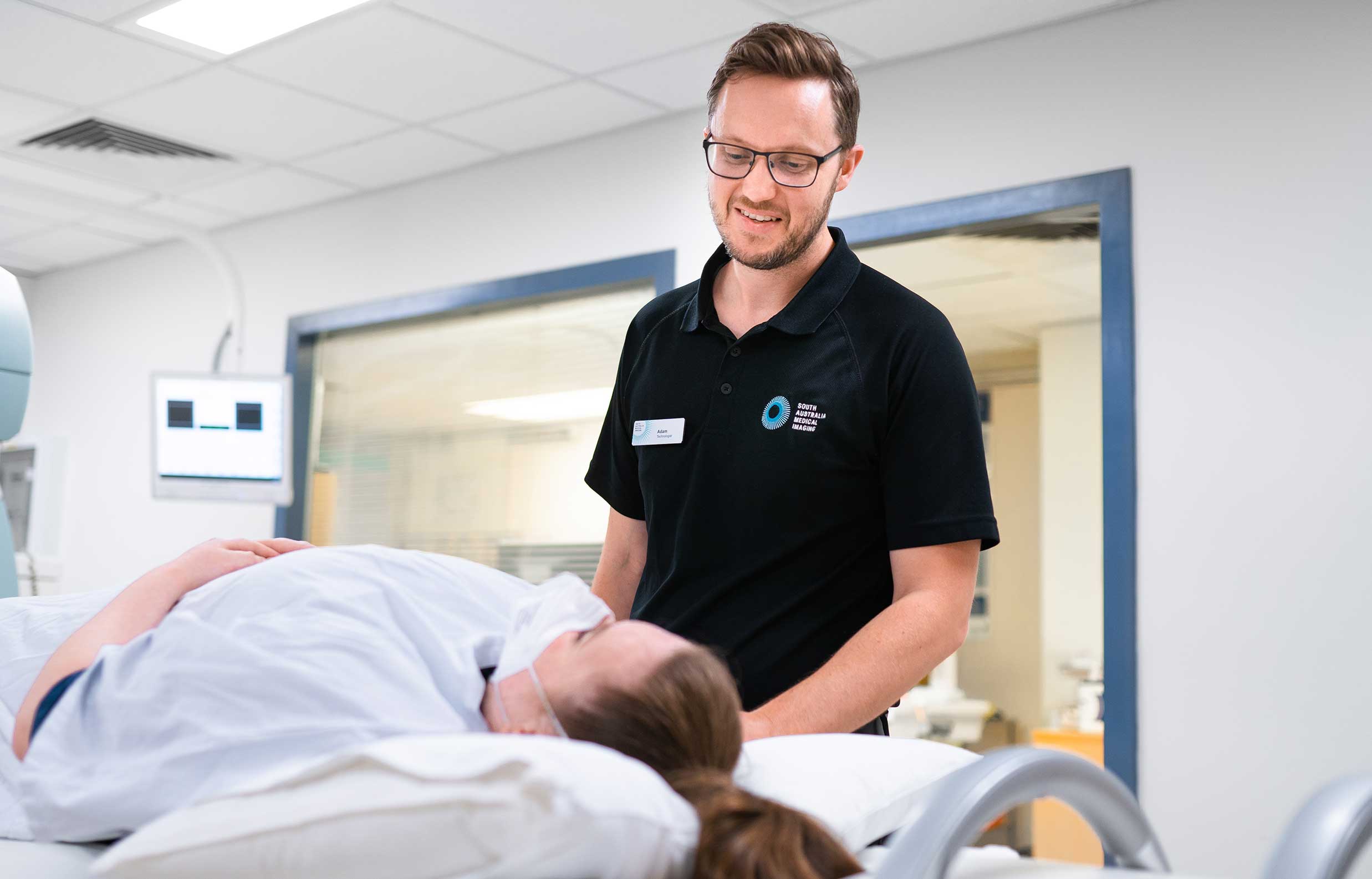
Nuclear medicine tests involve giving a patient a small amount of a radioactive tracer attached to a pharmaceutical compound, called a radiopharmaceutical. Following this, images are taken using a special scanner called a nuclear medicine gamma camera, which detects the distribution of the radiation being emitted from the body. These images allow assessment of different organs throughout the body depending on the type of radiopharmaceutical used.
The radioactive tracer is specific to the scan being performed and is most commonly injected into the blood stream through a vein, but might be given in different ways, including:
- swallowed
- injected directly into the tissue beneath the skin or
- inhaled (breathed in).
Only a small, amount of radiopharmaceutical is given to the patient to keep their radiation dose to a minimum whilst achieving good quality results.
Radioactivity can also be used as a therapy to treat some cancers or conditions. In these cases, the amount of radiopharmaceutical given is greater, and it mostly goes to (targets) the diseased tissue or organ. The type of radiopharmaceutical given emits ionising radiation that has the maximum effect on the part of the body or organ system being treated.
The incidence of allergic reaction to a radiopharmaceutical is very low and most do not stay in your body for long.
What is a bone scan?
A nuclear medicine bone scan can be used to detect bone injury (such as a broken bone), bone disease (such as cancer) or infection in the bones. It can also be used to review response to treatment.
There are two parts to a bone scan.
- a small amount of radiopharmaceutical is injected into the blood stream through a vein. Sometimes images are taken immediately after the injection to look at the blood flow to the area being scanned. Part 1 can take 15 to 30 minutes. Whether you have ‘early’ imaging will depend on why your doctor has requested the scan. The radiopharmaceutical attaches to the bone over time, and is imaged using a special scanner (gamma camera).
- after 2 to 4 hours, the patient returns to the department for ‘delayed’ images now that the radiopharmaceutical has made its way to the bone. These images can take 60 minutes.
There is no special preparation for a bone scan. It is recommended that the patient is well hydrated.
Before the start of the test, please inform the staff if you are pregnant or breastfeeding, or markedly claustrophobic.
What is a Myocardial Perfusion Scan (MPS)?
Also known as a nuclear medicine cardiac stress test.
This test looks at the blood supply to the heart muscle and provides information on how the heart is working.
To prepare for the test, please wear comfortable clothing and shoes that are suitable for light exercise.
It is important that you do not have a large meal before the test. All forms of beverages and foods containing caffeine (e.g. coffee, tea, cola and chocolate) should be avoided for 24 hours before the test.
There are some medications you might be taking that work by slowing the heart rate (e.g. beta blockers). These medications might need to be stopped before the stress test. Please discuss this with your doctor and the nuclear medicine team before stopping any medication.
There are two parts of a myocardial perfusion scan.
- Stress component: The stress component of the test is aimed at increasing the blood flow to the heart to show any changes in flow more clearly. The Nuclear Medicine specialist will need to assess your heart under “stress”. This can be done be exercising on a treadmill, or if you are unable to exercise sufficiently, a medication to mimic exercise will be used to increase blood flow to the heart.
When your heart has reached a target work capacity, a radiopharmaceutical (radioactive tracer) is injected into the blood stream through a vein. You will then be asked to rest in the waiting room for approximately 30 minutes. During this time, you might be given a drink of water, or a small tub of ice cream to eat. This helps make the images of your heart clearer. Occasionally you may experience side effects if a pharmaceutical is used to stress your heart. These side effects will be discussed with you by the nuclear medicine team prior to starting and will be treated if necessary.
After 30 minutes, the first set of images of the heart is obtained using a gamma camera. Images take approximately 20 minutes while the camera rotates around your heart. A CT scan at a low radiation dose, is also usually done as part of the study for further detail. After review of the images, you may be given an appointment time to return in the afternoon for Part 2 (usually 3–4 hours after Part 1).
- This part assesses your heart in its ‘resting’ state. You will be given a second injection of the same radiopharmaceutical. Again, you will be asked to sit in the waiting room for approximately 30 minutes. Your second set of images is then taken similar to your scan in the morning, in approximately the same time.
Before the start of the test, please inform the staff if you are pregnant or breastfeeding, or markedly claustrophobic.
What is a lung (VQ) scan?
A ventilation–perfusion (VQ) scan is a nuclear medicine scan that uses a radiopharmaceutical (radioactive tracer) to examine the airflow (ventilation) and blood flow (perfusion) in the lungs.
There are two parts to the scan:
- The radioactive tracer is inhaled to show the air flow to the lungs. Images are taken by a gamma camera which rotates around your chest for approximately 10 minutes.
- A radiopharmaceutical is injected into a vein in your arm. Similar images are again taken, for the same length of time. This part demonstrates the blood flow to the lungs.
In total the scan takes 30 to 45 minutes.
There is no special preparation for a lung scan and you won’t feel any differently.
Before the start of the test, please inform the staff if you are pregnant or breastfeeding, or markedly claustrophobic.
What is a renal scan?
A renal scan is a study which looks at the function of your kidneys. A small amount of radiopharmaceutical (radioactive tracer) is injected into the blood stream through a vein. Images are taken immediately after the injection using a gamma camera. Images show the blood flow to the kidneys, and the production and clearance of urine from the kidneys through the ureters to the bladder.
At the end of the scan, you may be asked to go to the toilet and empty your bladder, then return for a further 2 minutes of imaging.
The Nuclear Medicine Specialist may then decide to give a medication called Lasix (Furosemide) to encourage the kidneys to produce more urine. The kidneys are then scanned for a further 20 minutes.
The test will take approximately 30 to 60 minutes in total.
Please be well hydrated for the study.
Before the start of the test, please inform the staff if you are pregnant or breastfeeding, or markedly claustrophobic.